Cervical Facet Radiofrequency Neurotomy - Neck
Click the white PLAY button to start video.
Cervical facet radiofrequency neurotomy (facet rhizotomy) is used to treat nerve pain in the neck and/or shoulder. This technique is useful for those patients who experience short term relief following local anesthetic blocks of the nerves supplying the cervical facet joints. The procedure “turns off” the specific nerve that carries information about pain. The treatment can provide pain relief for about a year, but can last much longer for some people.
Read more about Cervical Facet Radiofrequency Neurotomy - Neck
Introduction
Cervical facet radiofrequency neurotomy (facet rhizotomy) is used to treat nerve pain in the neck and/or shoulder. This technique is useful for those patients who experience short term relief following local anesthetic blocks of the nerves supplying the cervical facet joints. The procedure “turns off” the specific nerve that carries information about pain. The treatment can provide pain relief for about a year, but can last much longer for some people.
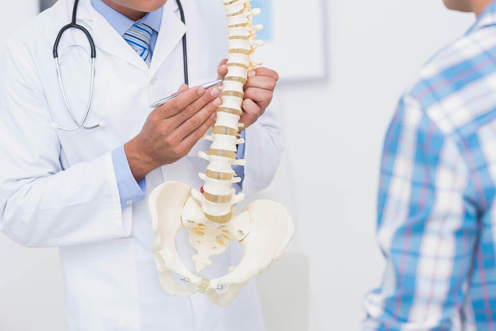
Anatomy
Your spinal cord is located within the protective spinal canal. The spinal cord extends from the brain and is a major part of your nervous system. Spinal nerves extend from the spinal cord and travel out of the spine to exchange nerve signals with your brain about specific parts of your body. In particular, medial nerves carry nerve signals about facet joint pain.
Causes
Symptoms
Facet joint disease causes pain in the neck that may spread to the back of the head, your shoulders, upper arms, and rarely the hands. Your neck may have powerful muscle spasms, so strong that the facet joints are moved out of position.
Diagnosis
Treatment
A cervical facet radiofrequency neurotomy is an outpatient procedure. You will wear a gown for the procedure and be positioned lying face down on a table. You will receive relaxation medicine before your procedure begins. The back of your neck will be sterilized and numbed with an anesthetic medication.
Your doctor will use a live X-ray image (fluoroscopy) to carefully insert and guide a needle-like tube (cannula) to the affected medial nerve. A small needle-like electrode (radiofrequency electrode) is inserted through the cannula. To ensure the cannula is in the correct position, a very mild electrical current is delivered through the electrode to the nerve. The nerve will briefly conduct pain signals and cause a muscle twitch, confirming that the correct nerve is targeted. Next, numbing medication is provided to the nerve in preparation for the treatment. Heat is delivered through the electrode to the nerve. The heat creates a lesion on the nerve. The heat disrupts the nerve’s ability to send signals about pain. At the end of the procedure, the cannula and electrode are removed. The process can be repeated for additional nerves that require treatment.
It usually takes three to four weeks for the treated nerves to completely die. During this period, your neck may feel weak. You may experience pain until the treated nerves are dead.
Cervical facet radiofrequency neurotomy typically results in pain relief for about 9 to 14 months or longer. About 50% of people experience pain relief for as much as two years. A small percentage of people do not experience any pain relief from the procedure. Over time, the nerves will grow back (regenerate). Some people will not experience pain again. If you experience pain, the procedure may be repeated.
Copyright © - iHealthSpot Interactive - www.iHealthSpot.com
This information is intended for educational and informational purposes only. It should not be used in place of an individual consultation or examination or replace the advice of your health care professional and should not be relied upon to determine diagnosis or course of treatment.
The iHealthSpot patient education library was written collaboratively by the iHealthSpot editorial team which includes Senior Medical Authors Dr. Mary Car-Blanchard, OTD/OTR/L and Valerie K. Clark, and the following editorial advisors: Steve Meadows, MD, Ernie F. Soto, DDS, Ronald J. Glatzer, MD, Jonathan Rosenberg, MD, Christopher M. Nolte, MD, David Applebaum, MD, Jonathan M. Tarrash, MD, and Paula Soto, RN/BSN. This content complies with the HONcode standard for trustworthy health information. The library commenced development on September 1, 2005 with the latest update/addition on February 16, 2022. For information on iHealthSpot’s other services including medical website design, visit www.iHealthSpot.com.